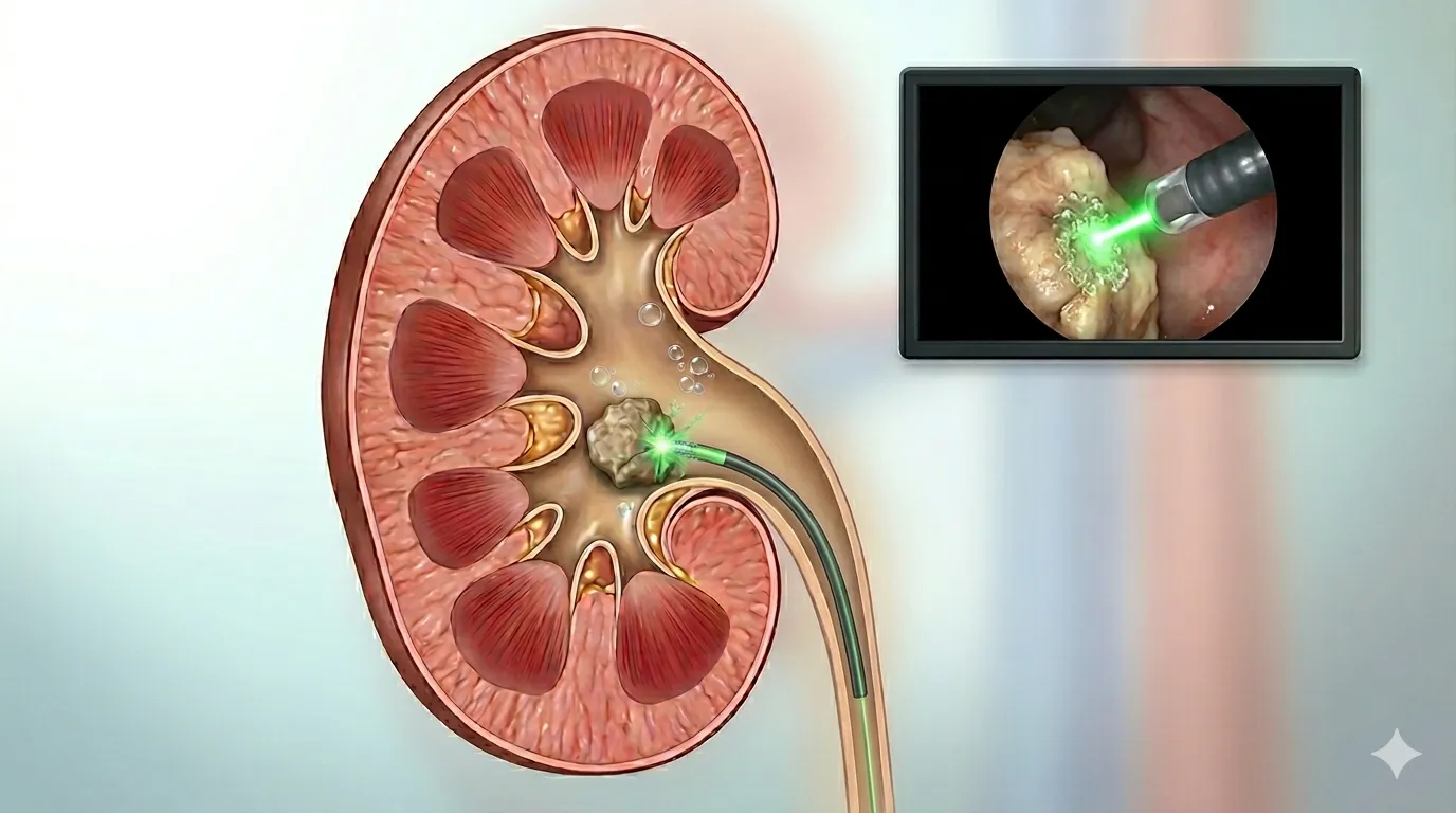
Renal colic is one of the most severe pains a person can experience. Patients describe it as a sharp, unbearable flank pain that radiates toward the groin and does not let up in any body position. The cause is a tiny, often only a few millimeters wide kidney stone or stone in the ureter — and precisely because of the severity and frequency of symptoms, this is one of the most common reasons for urgent urological care.
The good news: about 80% of smaller stones pass spontaneously, and for those that don't, modern urology offers minimally invasive techniques that have nearly eliminated the need for open surgery. The most effective method for stones up to 2 cm in the kidney is FURS with laser lithotripsy — no incision, with discharge home on day one or two.
This guide covers: why stones form, how to recognize the symptoms, what your urologist will do at the visit, and what all the treatment options are in 2026.
What is a kidney stone and how does it form?
The kidneys filter about 180 liters of plasma daily and excrete around 1.5–2 liters of urine. Urine contains dissolved salts — primarily calcium, oxalate, uric acid, phosphate and citrate. When the concentration of these salts exceeds their solubility (due to dehydration, diet, metabolic disorders), they begin to crystallize. The crystals slowly merge and grow, forming a stone.
Key point: stones don't form overnight. Tiny crystals accumulate in the kidney over decades, most often in the renal papilla. When a stone grows and breaks loose, it enters the collecting system and can pass down the ureter toward the bladder. That is when symptoms begin.
Stone types
- Calcium oxalate (about 70–80%) — the most common. Linked to salt and oxalate intake, and obesity.
- Calcium phosphate (5–10%) — often associated with alkaline urine and parathyroid disorders.
- Uric acid (5–10%) — in patients with gout, diabetes and acidic urine; can be dissolved by alkalinizing the urine.
- Struvite (5–10%) — form in the presence of chronic infections; grow rapidly and can fill the entire collecting system (staghorn stone).
- Cystine (less than 1%) — hereditary; patients have recurrences from a young age.
The type of stone matters because it determines the prevention strategy. After removal, the stone is always sent for chemical analysis.
How common are kidney stones?
- Lifetime risk of forming a kidney stone: 10–15% in men, 5–8% in women.
- Incidence in Europe is rising — due to obesity, high-salt diets and climate change.
- Without prevention, recurrence within the first 5 years is around 50%. With prevention, it falls to 10–15%.
- The average age at first stone is 30–50, but stones also occur in children.
Symptoms — how to recognize a stone
A small stone that stays in the kidney often causes no symptoms and is discovered incidentally (e.g. on ultrasound done for another reason). Symptoms appear once the stone moves down the ureter:
1. Renal colic
A typical sharp pain that starts in the flank and radiates forward toward the groin — in men further toward the scrotum, in women toward the labia. The pain is called colic — it comes and goes in waves of 20–60 minutes. The patient cannot find a position to ease the pain, often pacing, bending over or seeking a hot bath.
2. Hematuria (blood in the urine)
Microscopic (visible only in the lab) or macroscopic (urine appears pink, red or tea-colored). The blood comes from injury to the ureteral lining as the stone passes.
3. Nausea and vomiting
A reflex response associated with severe pain. Often even fluids cannot be kept down, worsening dehydration and the situation overall.
4. Urinary symptoms
When the stone reaches near the bladder, frequent and painful urination, urgency and a sense of incomplete emptying appear — similar to a urinary tract infection.
5. Fever — a red flag
If a stone is accompanied by a temperature above 38°C, chills and flank pain — this can indicate infection above an obstruction (obstructive pyelonephritis or urosepsis). This is a medical emergency and requires urgent placement of a JJ stent or nephrostomy.
When to see a urologist urgently
Go to the emergency department or contact us immediately if you have:
- Renal colic with a temperature above 38°C or chills.
- Pain that does not stop despite medication.
- Blood in the urine with flank pain.
- Anuria (complete absence of urine) — especially if you have only one functional kidney.
- A stone in pregnancy — always managed in a multidisciplinary setting.
How is the diagnosis made?
History and physical examination
A detailed discussion about the pain, prior stones, medications, diet and hydration. Examination of the abdomen and percussion over the flank (positive in obstruction).
Blood and urine tests
- Urinalysis — blood, crystals, infection (leukocytes, nitrites). Urine culture if infection is suspected.
- Blood — creatinine (kidney function), calcium, uric acid, CRP (when infection is suspected).
Imaging
- Kidney and bladder ultrasound — first choice. Detects a stone in the kidney (>3 mm), pelvic dilatation (hydronephrosis) and a stone in the distal ureter near the bladder. Ultrasound does not reliably detect stones in the mid-ureter.
- NCCT (non-contrast CT — "stone protocol") — the gold standard. It detects all stone types, gives accurate size, location and density (Hounsfield units — useful for predicting ESWL success). The radiation dose is significantly lower than for a standard CT.
- Plain abdominal X-ray (KUB) — less accurate, used less frequently.
- IVU (intravenous urography) — largely replaced by CT, but still useful for specific anatomical questions.
Metabolic workup
Indicated for recurrent stones, first stone in a child, large or staghorn stones, or in patients with comorbidities. It includes a 24-hour urine collection (calcium, oxalate, uric acid, citrate, volume) — showing exactly what to correct in prevention.
Treatment options for kidney stones
The choice of method depends on several factors: stone size, location, density, kidney anatomy, presence of obstruction or infection, and the patient's overall health. There is a clear hierarchy from least to most invasive:
1. Conservative
For asymptomatic kidney stones smaller than 5 mm, and for ureteral stones smaller than 5–6 mm without signs of infection or kidney damage — observation for up to 4 weeks is recommended, supported by:
- Hydration — 2.5–3 L of water per day.
- Alpha-blockers (tamsulosin, silodosin) — relax the lower ureter and speed up stone passage ("Medical expulsive therapy — MET").
- Analgesics — diclofenac or ibuprofen for pain control; opioids for severe colic.
- Antiemetics — when nausea is prominent.
2. ESWL (extracorporeal shock-wave lithotripsy)
The stone is broken up by shock waves generated outside the body and focused on the stone. No incision, usually no anesthesia (only sedation). Indicated for kidney stones < 2 cm and proximal ureteral stones < 1 cm with density below ~1000 HU.
Limitations: ineffective for dense stones (cystine, brushite, some calcium oxalate), in patients on anticoagulants, and in patients with anatomical variants; success drops considerably for stones in the lower pole of the kidney.
3. URS and FURS (ureteroscopy)
Endoscopic access through the urethra and bladder, using a laser to fragment the stone.
- Semi-rigid URS — a rigid instrument; ideal for stones in the distal and mid-ureter.
- FURS (flexible ureterorenoscopy) — a flexible, thin instrument that reaches the kidney through natural orifices. With a holmium or thulium laser, the stone is dusted or extracted with a basket. The most effective minimally invasive method for kidney stones up to 2 cm. Details in the FURS guide.
A JJ stent is often placed for 1–4 weeks after FURS — preventing obstruction caused by ureteral swelling. It is removed in an outpatient flexible cystoscopy. Detailed guide: JJ stent — why it is placed, how long it stays and how it is removed.
4. PCNL (percutaneous nephrolithotomy)
Direct access to the stone through a small incision (about 1 cm) in the back — a narrow tract is created from skin to kidney with a nephroscope. The stone is broken up with a laser or ultrasound and the fragments are removed. The most effective method for large stones (> 2 cm), staghorn stones and complex anatomies. Hospital stay 2–4 days.
Mini-PCNL and ultra-mini-PCNL use smaller instruments (3–5 mm), reducing bleeding and speeding recovery; their use is expanding.
5. Combined procedures
For particularly complex cases, PCNL + FURS are combined in the same operation or in two stages — known as "endoscopic combined intrarenal surgery" (ECIRS).
6. Open surgery
Today extremely rare — less than 1% of stones. Indicated only for extremely complex anatomical variants or combined operations for other diseases.
Complications of an untreated stone
- Hydronephrosis and kidney damage — prolonged obstruction gradually impairs kidney function; irreversible after several weeks of complete blockage.
- Obstructive pyelonephritis and urosepsis — the most dangerous complication; mortality without prompt treatment is significant.
- Anuria — when there is a stone in a single functional kidney or simultaneously in both ureters.
- Formation of new stones — stasis and infection promote accelerated growth.
Prevention — how to avoid a new stone
Prevention is so important that every patient after a first stone should go through at least a minimal prevention program. For recurrent stones, a metabolic workup and targeted therapy are performed.
Universal recommendations (for all stone types)
- Fluids — at least 2.5–3 L per day (target: 2 L of urine per day). Watch the color of your urine — it should be pale yellow.
- Salt — less than 5 g per day. High salt intake increases calcium excretion.
- Animal protein — moderate; too much meat increases calcium and uric acid excretion and decreases citrate.
- Body weight — obesity is a risk factor.
- Lemon and citrate — citrate inhibits stone formation; lemon juice diluted in water is a simple and effective measure.
Specific to stone type
- Calcium oxalate — do not reduce dietary calcium (paradoxically increases risk); moderate oxalates (spinach, chocolate, nuts, tea).
- Uric acid — alkalinize the urine (potassium citrate), reduce purines (red meat, organ meats, alcohol), control uric acid (allopurinol in patients with gout).
- Struvite — eradicate chronic urinary infections; often requires complete stone removal.
- Cystine — high hydration, alkalinization, specific medications (tiopronin).
Frequently asked questions
How painful is a kidney stone?
Renal colic is considered one of the most severe pains in medicine — often described as worse than childbirth. The pain is sharp and colicky, and the patient cannot find a comfortable position. The good news: it is well controlled with analgesics and disappears as soon as the stone passes the narrowest part of the ureter.
Does every stone need an intervention?
No. Stones up to 5 mm pass spontaneously in about 80% of cases. Small asymptomatic stones in the kidney can be observed for years. Intervention is needed for stones causing obstruction, infection, severe pain, or those too large to pass on their own.
Which is the best method — FURS, PCNL or ESWL?
There is no single "best" method — there is a best method for your situation. ESWL is the least invasive but has the lowest success rate. FURS is optimal for kidney stones up to 2 cm. PCNL is irreplaceable for stones over 2 cm. Based on CT, location and stone density, the urologist chooses the method with the best balance of success and risk.
Does FURS hurt?
The procedure itself is performed under general or spinal anesthesia — you feel nothing during it. After the procedure, the most common complaints are mild flank tenderness and a sense of irritation when urinating (because of the JJ stent if placed). All of this usually resolves within a few days with mild analgesics.
What is a JJ stent and is it noticeable?
The JJ stent is a thin silicone tube that keeps the ureter open. Most patients feel it as a mild discomfort when urinating and occasional flank pressure — all of this is expected and temporary. The stent is removed in 1–4 weeks via flexible cystoscopy.
Can I fly with a kidney stone?
With a small asymptomatic stone in the kidney — yes. But with active colic or an obstructing stone in the ureter, travel should be postponed. Pressure changes on a plane do not cause colic, but they make matters worse if colic develops at altitude where there is no medical assistance.
Does mineral water cause stones?
No. On the contrary — sparkling mineral water can help (it contains citrate). What causes stones is insufficient overall fluid intake and a diet rich in salt and animal protein — not the type of water you drink.
Next step — from urinary colic to a lasting solution
If you've had renal colic, if an ultrasound has shown a stone, or if you have recurrent symptoms — the best time for an examination is now. The goal isn't only to remove the current stone, but to understand why it appeared and build prevention so it doesn't return.
At the visit we'll go through your symptoms together, do an ultrasound and lab work, arrange a CT if needed, and create a treatment plan. Book a urological appointment — by phone or contact form.
For international patients: we organize diagnostics and the FURS procedure in a single trip, with full follow-up instructions in English for your local doctor.